Figure 3.
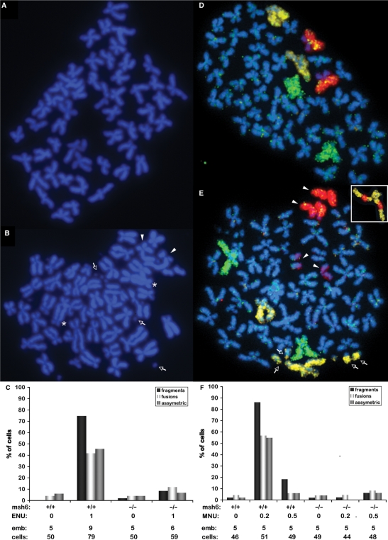
Alkylation-induced lethality is caused by MMR-dependent chromosomal aberrations. (A) Metaphase spread of an untreated embryo showing the 50 regular chromosomes of a single cell. (B) Metaphase spread of a typical cell from a wild-type embryo treated with 1 mM ENU, showing chromosome fragments (arrows), fused chromosomes (asterisks), and asymmetric chromosomes (arrowheads). (C) Quantitation of the three types of aberrant chromosomes mentioned in (B). Below the graph are indicated the msh6 genotype, ENU treatment (mM), number of embryos scored, and total number of cells scored. (D) Metaphase spread from an untreated embryo with chromosome paints for chromosomes 4 (yellow), 5 (green) and 7 (red), showing two of each chromosome per cell. (E) Metaphase spread of a typical cell from a wild-type embryo treated with 1 mM ENU, stained with chromosome paints for chromosome 4 (yellow), 5 (green) and 7 (red); indicated aberrations include fragments of chromosome 4 (arrows), and both chromosomes 7 having broken into halves (arrowheads) [note that in both (D) and (E) the short arms of chromosome 7 show weaker staining than the long arms]. Inset in (E): example of a rearranged chromosome 4 with fragments of chromosome 7. (F) Quantitation of the three types of aberrant chromosomes mentioned in B after MNU exposure. Below the graph are indicated the msh6 genotype, MNU treatment (mM), number of embryos scored, and total number of cells scored.

