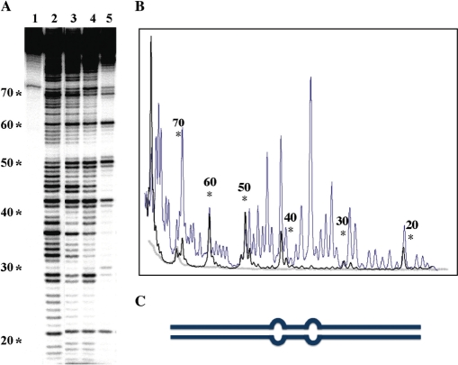
Figure 7.
TmHU condenses DNA. (A) Two-dimensional DNase I footprinting with 89 bp looped DNA. The position of loops corresponds to bases 40–41 and 49–50 (positions are identified to the left of the sequencing gel). Reactions in (A) were: lane 1, DNA only; lane 2, DNA treated with DNase I in absence of TmHU; lanes 3–5 TmHU:DNA complexes 1, 2 and 3, respectively, as identified in (A). All reactions were subjected to EMSA and either free DNA (lanes 1 and 2) or TmHU–DNA complex (lanes 3–5) isolated from the gel as described in Materials and methods section prior to denaturing gel electrophoresis to separate cleavage products. (B) Densitometry profile of uncut DNA (gray line), DNase I-digested DNA (blue) and TmHU:DNA complex 3 (black). (C) Cartoon illustration of the 89 bp looped DNA.

