
Figure 4.
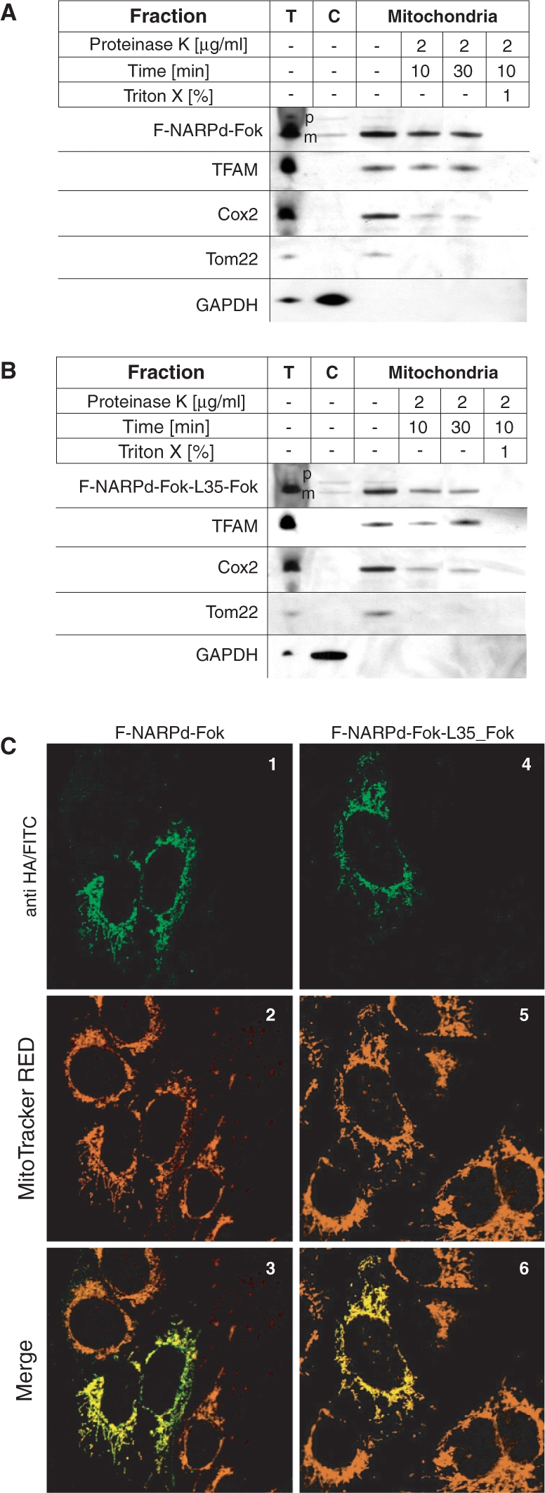
ZFNs are localized in mitochondria in cells. (A) and (B) The F-NARPd-Fok and F-NARPd-Fok-L35-Fok ZNFs localize inside mitochondria. The 143B cells harbouring wt mtDNA were transiently transfected with monomeric F-NARPd-Fok (A) or single-chain ZFN—F-NARPd-Fok-L35-Fok (B), fractionated 48 h post-transfection and the protein fractions were analysed by western blotting using anti-HA mAb. The localization of the ZFN precursors (‘p’) and their mature (‘m’) form in total cell lysate (‘T’), cytosolic (‘C’) and a mitochondrial fraction treated with proteinase K under various conditions as indicated, was compared with the localization of marker proteins. The precursors of mitochondrial ZFNs, found in the mitochondrial fractions, were located outside the mitochondria, since they were accessible to protease digestion. In contrast, the mature form of ZFNs was protected and became accessible to proteolysis only after the mitochondria were lysed with Triton X-100. The following endogenous proteins were used as fractionation markers: (i) TFAM: the transcription factor that is localized in the mitochondrial matrix; (ii) Cox2; a subunit of the cytochrome oxidase complex localized in the mitochondrial inner membrane; (iii) Tom22: a subunit of mitochondrial translocase of outer membrane; and (iv) GAPDH: a protein localized in cytoplasm. (C) The F-NARPd-Fok and F-NARPd-Fok-L35-Fok ZFNs co-localize with mitochondria. The intra-cellular localization of ZFNs was additionally analysed by immunofluorescence in transiently transfected 143B cells. ZFNs were detected with antibodies against the HA epitope-tag followed by secondary antibodies conjugated to FITC (1 and 4; green) Mitochondria were stained with MitoTracker CMX Red (2 and 5; red). Both ZFNs exhibited mitochondrial-staining pattern that in represented by yellow staining on digitally overlaid pictures (3 and 6).