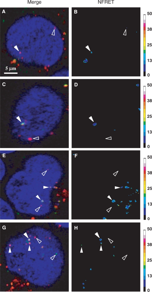
Figure 3.
FRET imaging of double-labelled polyplexes in living cells. C2C12 (A–D) and HeLa (E–H) cells were transfected for 5 h with fluo-p3NF-luc-3NF/rho-LPEI (A, B and E, F) or fluo-p3NF-luc-3NF/rho-HIS (C, D and G, H). NFRET ratio images were calculated according to Xia's normalization. Fluo, rho and Draq5 were excited at 488, 543 and 633 nm, respectively. Sidebars: colour pixels scoring for FRET level from 0 (dark blue) to 50 (white). Each image corresponds to 512 × 512 pixels measuring 0.28 × 0.28 µm2 each.

