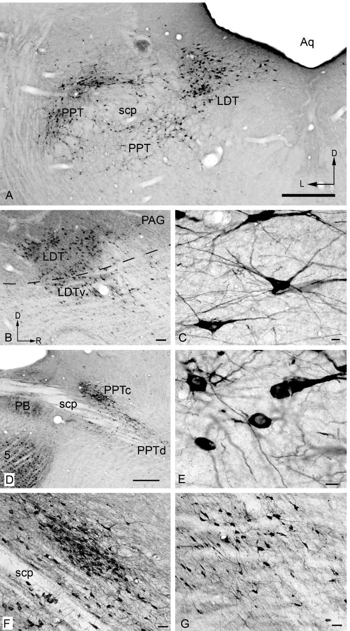
Figure 4.

Photomicrographs showing ChAT-IR cells in traditionally-named cholinergic nuclei. A) pedunculopontine (PPT) and laterodorsal tegmental (LDT) nuclei (compare with Fig. 2, approx. section 16). D - Dorsal; L - lateral. B) Parasagittal section showing LDT and its ventral subdivision (LDTv), located ventral to the border of the periaqueductal gray (dotted line). Dorsal is up and rostral to the right in this and subsequent panels. C) LDT cells. D) Parasagittal section showing the relationship between the parabrachial nuclei (PB), the compact part of PPT (PPTc) and the dissipated part of PPT PPTd). E) PPTd cells. F) compact PPT. G) dissipated PPT. Scale bar = 0.5 mm for A, D; 100 μm for B; 10 μm for C, E; and 50 μm for F, G.
