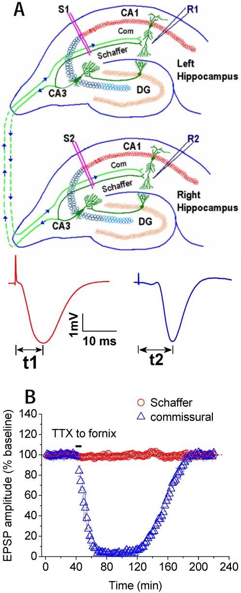
Figure 1. Schaffer and commissural pathways in CA3-CA1 network.
A, Recording electrode (R1) in CA1 stratum radiatum records field excitatory postsynaptic potentials (fEPSPs) in response to stimulation of ipsilateral Schaffer (S1) and contralateral commissural pathway (S2) at stratum radiatum, respectively. Schaffer = Schaffer collaterals; Com = commissural fibers. B, Peak latency of Schaffer fEPSP (t1) is about 5 ms shorter than that of commissural fEPSP (t2). Timing window (Δt) between Schaffer and commissural inputs is calculated from the inter-peak intervals (t2−t1). Infusing tetrodotoxin (TTX), the sodium channel blocker, bilaterally into the fornix where commissural fibers cross the midline suppressed the fEPSP from commissural pathway, but had no effect on the fEPSP from Schaffer pathway (n = 4).

