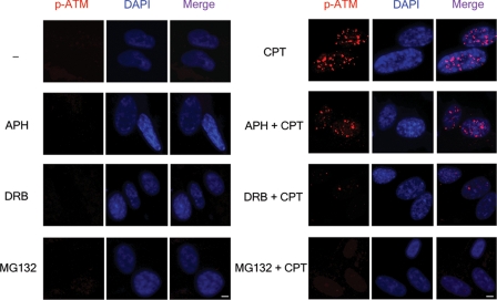
FIGURE 3.
In situ visualization of autophosphorylated ATM foci in CPT-treated quiescent WI-38 cells. WI-38 cells cultured in 0.2% serum were pretreated with DMSO (0.1%), APH (10 μm), DRB (150 μm), or MG132 (2 μm) for 30 min, followed by 1-h co-treatment with CPT (25 μm). The cells were fixed by 3.7% paraformaldehyde and stained with both anti-p-Ser-1981-ATM antibody and 4′,6′-diamino-2-phenylindole (DAPI). Scale bar, 5 μm.

