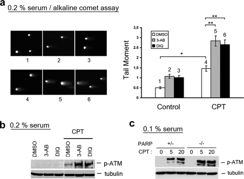
FIGURE 6.
Inactivation of PARP1 elevates the amount of CPT-induced SSBs and the level of ATM autophosphorylation. WI-38 cells cultured in DMEM supplemented with 0.2% serum were pretreated with 4 mm 3-AB or 100 μm DIQ for 30 min, followed by co-treatment with CPT (25 μm) for 1 h. The cells were subjected to either alkaline comet assay (see representative comet images to the left and the histogram of the tail moment to the right; p values for comparisons marked * and ** were < 0.005 as determined by two-tailed Student's t test) (a) or immunoblotting with indicated antibodies (b). c, parp+/- and parp-/- primary MEFs cultured in 0.1% serum (for 3 days) were treated with CPT for 1 h, followed by immunoblotting with antibodies as indicated. DMSO, dimethyl sulfoxide.

