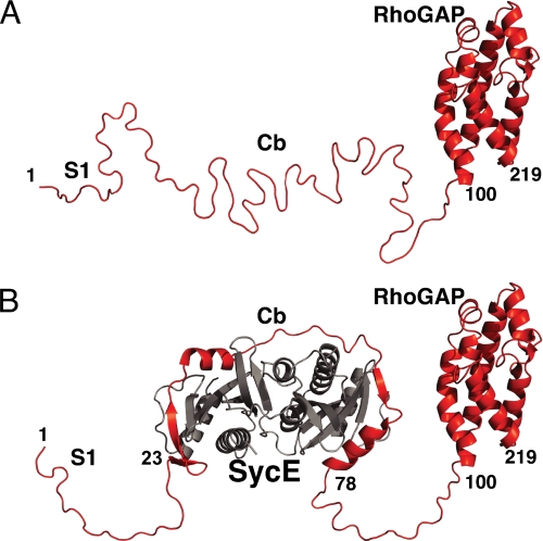
FIGURE 5.
Free and SycE-bound YopE. A, a ribbon representation of free YopE, with the crystal structure of the RhoGAP domain (23) depicted and other portions in modeled random coil conformation. B, a ribbon representation of the SycE-YopE complex, with the conformation of SycE (gray) and the YopE Cb region corresponding to the crystal structure of SycE-YopE(Cb) (6) and the conformation of the YopE RhoGAP domain corresponding to its crystal structure (23); other portions are depicted in modeled random coil conformation.

