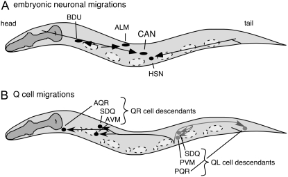
Figure 1.—
C. elegans cell migrations. Anterior is to the left and dorsal is up in all figures. (A) Embryonic cell migrations. Schematic lateral view of a newly hatched first larval stage hermaphrodite. Both the final positions of the ALM, BDU, CAN, and HSN cell bodies (ovals and circle) and their migration routes (arrows) are indicated. Dashed ovals show the positions of landmark nuclei (V cells) used in assessing cell position. (B) Q-neuroblast migrations. Schematic lateral view of first larval stage animal after the Q descendants have completed their migrations. Indicated are the final positions of the QR descendants SDQ, AVM, and AQR (solid circles) and their migration routes (solid arrows) and of the QL descendants SDQ, PVM, and PQR (shaded circles) and their migration routes (shaded arrows). Cell divisions and cell deaths in the Q lineages are not shown. Dashed ovals and circles show locations of the landmark hypodermal nuclei, Vn.a and Vn.p, used in assessing cell position.

