Figure 3.
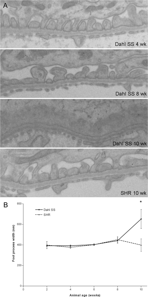
Electron microscopy of podocytes in Dahl SS and SHR rats (A), and evaluation of the mean foot process width (B). In 4-week-old Dahl SS rats (a, top panel), the podocyte ultrastructure was normal, showing regularly spaced foot processes. Occasionally, protein droplets were observed in podocyte major processes and cell bodies (arrowheads). At 8 weeks, subtle coarsening of the foot processes was observed sporadically in Dahl SS rats (A, second panel). At 10 weeks of age, widespread foot process effacement was observed in segmental areas of the glomerulus, with condensation of the actin cytoskeleton at the basal site of the effaced processes (arrow). Microvillous transformation of podocytes was observed frequently at this time point. In contrast, the podocytes of SHR rats had normal ultrastructure throughout the time course studied (A, bottom panel). Original magnification: ×15,000. Quantification of the mean foot process width showed significant effacement only in the 10-week-old Dahl SS rats (B). An asterisk indicates P < 0.05.

