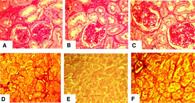
Figure 9.
Kidney and heart histology, chronic study. A–C: Periodic acid-Schiff-stained kidney cortical sections at 26 months of age (n = 5 per group). A: Reg: moderate glomerular and tubulointerstitial sclerosing changes. B: CR: scattered areas of interstitial fibrosis and tubular atrophy. The glomeruli were of normal size, but had recognizable sclerotic changes. C: CR-high: enlarged glomeruli and diffuse glomerular sclerosis, with tubular and interstitial changes more marked than CR mice. D–F: Sirius Red-stained coronal mid-ventricular sections (n = 5 mice/group), at 26 months. D: Reg: bands of connective tissue, concentrated near blood vessels; E: CR: no increase in connective tissue; F: CR-high: diffuse increase in connective tissue bands in the perivascular regions and fine bundles that surround irregularly enlarged bundles of myocytes. Original magnifications, ×250.

