Figure 5.
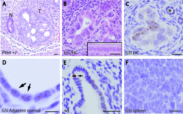
Mouse model of TII precancers. A and B: H&E stains; C–F, pH2AX IHC counterstained with hematoxylin. A: Endometrium from Pten+/− female (6 weeks) showing spontaneous endometrial lesion illustrating TI histology. T, Neoplastic focus; N, adjacent morphologically normal gland. B: Endometrium from fifth generation mTerc−/− (G5i) mouse (14 months). Inset shows morphologically normal surface epithelium from control G0i mouse (15 months). Note that EIC lesion is characterized by extreme nuclear anisometry and mild architectural abnormalities, whereas normal epithelium consists of well-organized, polarized columnar epithelium. C: pH2AX IHC from same G5i uterus in B shows many nuclear foci consistent with DSBs; asterisk indicates morphologically normal gland with small, nonatypical and isometric nuclei. D: Numerous pH2AX foci in adjacent normal epithelium. E: pH2AX in control G0i mouse shows occasional apoptotic cell (arrow) and rare pH2AX foci that are difficult to discern and far rarer than in G5i uteri. F: Spleen from G5i animal showing absence of pH2AX foci. Scale bars: 25 μm (A–C, E); 10 μm (D, F).

