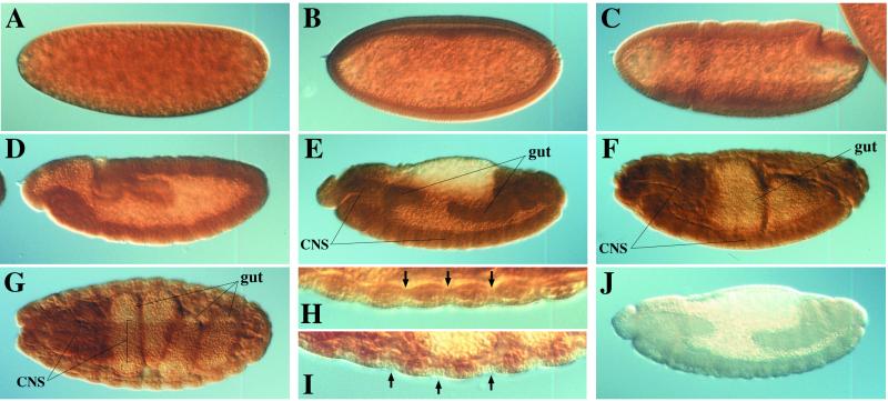
Figure 3.
Localization of DFak56 protein during embryogenesis. Polyclonal anti-serum 1562C, raised against the C terminus of DFak56, was used to stain fixed embryos: stage 3 (A), 5 (B), 6 (C), 11 (D), 13 (E), 15 (F), and 16 (G, seen from the ventral side). Magnified ventral views of stage 16 embryos are shown in H and I; arrows indicate muscle attachment sites (H) and ectodermal cells located at segmental junctions (I). Preimmune serum gave little or no signal (J).

