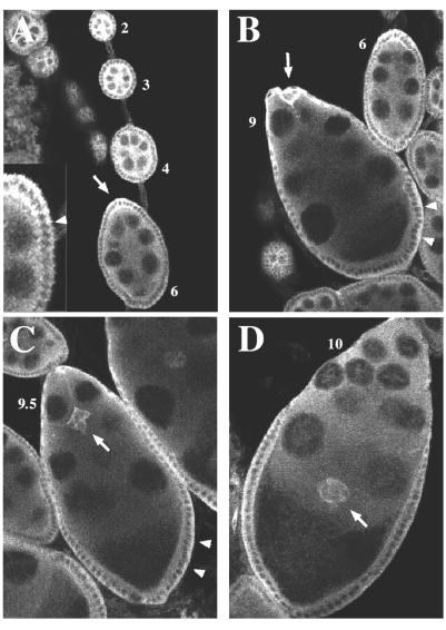
Figure 4.
Confocal images of egg chambers stained for DFak56 protein. (A) Stage 2 to stage 6 egg chambers (see numbers). The arrow indicates the anterior tip of the stage six egg chamber, and the inset shows an enlarged view of the right side of the anterior tip, slightly rotated. The arrowhead marks the boundary between high and low expression. (B) At early stage 9, border cells (arrow) begin to migrate between the nurse cells toward the oocyte at the posterior end of the egg chamber. Their migration continues through mid-stage 9 (C), and they reach the oocyte during stage 10 (D). Arrowheads in B and C indicate follicle cells at the posterior ends of the egg chambers. Control egg chambers stained with preimmune serum show a much lower, uniform signal (not shown).

