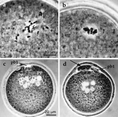
Figure 2.
The behavior of nuclear material from small or large ES cells after microinjection into enucleated mouse oocytes. (a) Small cell nuclear components condense to form a disorganized chromosome array 3–4 h after microinjection. (b) Large cell nuclear components condense to form an orderly chromosome array (resembling the maternal metaphase plate) 3–4 h after microinjection. (c) A one-cell embryo formed by the microinjection of a small E14 cell nucleus into an enucleated oocyte and activation by exposure to Sr2+ for 6 h in the presence of cytochalasin B. Two pseudo-pronuclei (pp) are discernible, each containing several nucleoli; remnants of the first polar body (pb1) are also visible. (d) A one-cell embryo formed by the microinjection of a large E14 cell nucleus into an enucleated oocyte and activation by exposure to Sr2+ for 6 h in the absence of cytochalasin B. A degenerate first polar body (pb1), single pseudo-pronucleus (pp), and pseudo-second polar body (indicated by an arrow) are visible.

