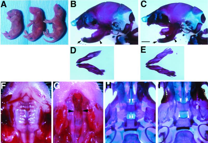
Figure 3.
Cleft palate in Lhx8 knockout mice. (A) Two dead newborn Lhx8 homozygous mice (Left and Center) are compared with their wild-type littermate (Right). (B and C) Skeletal staining of the head of a newborn Lhx8 homozygous mutant with cleft palate (C) as compared with that of a wild-type control (B), viewed from the side. The craniofacial features of the mutant mouse with cleft palate appeared grossly normal. In the mutant, the mandible was lower because the mouth was more widely open when the animal was fixed. Arrows and arrowheads point at the tip and the middle part of the mandibles in both the wild-type (B) and the mutant (C) animals, respectively. (D and E) Top view of the mandible of a newborn Lhx8 mutant animal with cleft palate (E) as compared with that of a wild-type control (D). The mandible of the mutant appeared normal. (F and G) Ventral view of the upper jaw of a newborn Lhx8 homozygous mutant (G) as compared with that of a wild-type control (F). Notice a cleft of the secondary palate (indicated by arrowheads). (H and I) Ventral view of the base of skulls of newborn wild-type (H) and Lhx8 homozygous mutant (I) mice stained with alcian blue and alizarin red. The palatal bones in the mutant failed to grow toward the midline (indicated by arrowheads). The scale bar in C represents 6.6 mm for panel A and 1.3 mm for panels B–-I.

