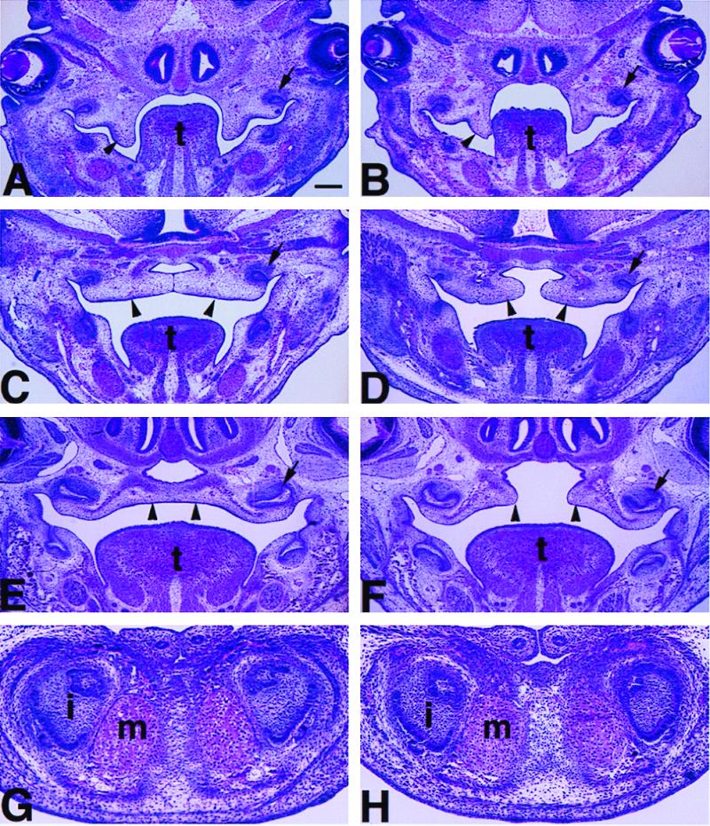
Figure 4.

Palate shelves elevated but failed to make contact and fuse in Lhx8 homozygous mutant mouse without detectable defects in the mandible. H&E staining of coronal sections through the palatal region of the homozygous mutant embryos (B, D, and F) at different developmental stages as compared with that of wild-type controls (A, C, and E). (A and B) E13.5; (C and D) E14.5; (E and F) E16.5. Arrowheads point at the palatal shelves that were not in contact (A, B, D, and F), were in contact (C), or were completely fused (E). Arrows point at one of the upper molar tooth buds in all panels. (G and H) H&E-stained coronal sections through the distal region of the mandible of a wild-type (G) and an Lhx8 homozygous mutant embryo with cleft palate at E16.5, showing normal development of the mandible, the incisor teeth (i), and the Meckel's cartilage (m) in the mutant. Scale bar in A represents 200 μm for panels A–F and 80 μm for panels G–H. t, tongue.
