Figure 1.
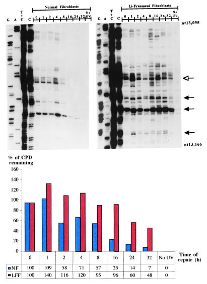
Repair of UVB-induced CPDs at nucleotide resolution along the NTS of the human p53 tumor-suppressor gene (exon 5, nucleotides 13,095–13,166) in normal human skin fibroblasts (Left) and LF041 skin fibroblasts (Right). The first four lanes from the left on each autoradiogram show LMPCR of DNA treated in standard Maxam–Gilbert cleavage reactions. The following eight lanes show LMPCR of DNA isolated from UVB-irradiated cells that have undergone repair for the indicated times (hr). The far right lane shows LMPCR of unirradiated DNA followed by T4 endonuclease V/photolyase digestion. The shaded arrows indicate dipyrimidine sites quantified with a Fuji BAS 1000 phosphorimager, equipped with the MacBAS v2.5 program. The open arrow indicates a representative dipyrimidine site (i.e., CC site indicated in bold, located within 5′-CCCCCG-3′, codon 151–152) for which the relative repair rate has been graphically illustrated below the autoradiograms. NF, normal fibroblasts; LFF, LF fibroblasts.

