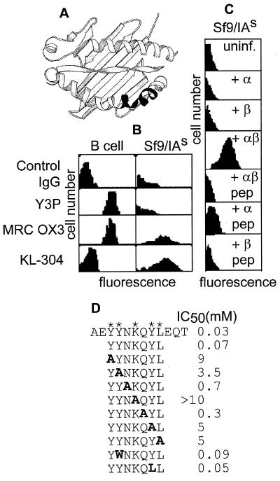
Figure 1.
KL-304 reactivity with IAs from human and insect cells. (A) The KL-304 epitope (23) (Lower Right, shaded) on a ribbon diagram of the class II MHC peptide-binding site (36). (B) Flow cytometry of IAs/d B cells (B-cell lymphoma LS 102.9) or IAs-expressing insect cells (Sf9/IAs), by using control and MHC-specific antibodies, as indicated (Left). KL-304 preferentially stains recombinant insect cells, as compared with B cells. Y3P, MRC OX3, and other antibodies tested (10–2.16, MRC OX6, 10–3.6.2, MK-S4; not shown) preferentially stain B cells or do not discriminate. (C) Flow cytometry by using KL-304 of insect cells expressing individual IAs subunits α, β, or both, with or without antigenic peptide PLP[139–151] treatment. Only the empty αβ complex binds KL-304. (D) KL-304 epitope mapping. Substitutions of the IAs sequence shown in bold, with asterisks indicating positions of key residues.

