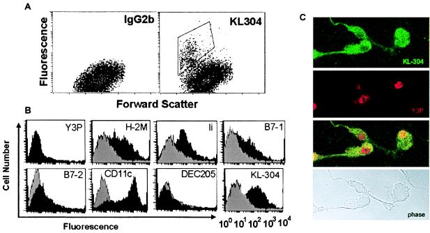
Figure 1.
KL-304-positive DC express surface markers consistent with immature DC. Bone marrow DC from SJL mice express peptide-receptive class II MHC molecules at the cell surface. (A) Staining with KL-304, specific for empty or peptide-receptive I-As, relative to an isotype-matched control antibody. (B) The KL-304-positive population sorted and analyzed for DC surface markers. (C) Confocal microscopy of DC doubly stained with FITC-labeled KL-304 and biotinylated Y3P/Texas red-labeled streptavidin. Shown are fluorescence images (optical magnification × 100) for KL-304, Y3P, costaining, and a phase-contrast image of the same field (top to bottom).

