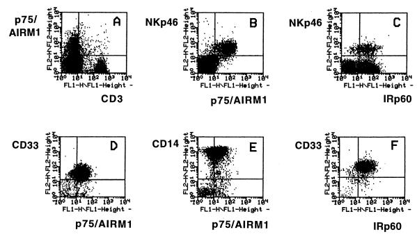
Figure 1.
Pattern of expression of p75/AIRM1 molecule in cord blood-derived lymphoid or myeloid cell populations. Cells were isolated from cord blood by the use of a F/H gradient and analyzed by two-color immunofluorescence and FACS analysis for the expression of p75/AIRM1 in combination with CD3, NKp46, CD14, or CD33 molecules. In A–C, analysis was performed on cells gated on lymphoid populations, whereas, in D–F, analysis was performed on cells gated on myeloid populations. C and F show lymphoid or myeloid cell populations stained with anti-IRp60 mAb in combination with anti-NKp46 or anti-CD33, respectively. The dot plots were divided into quadrants representing unstained cells (lower left), cells with only red fluorescence (upper left), cells with red and green fluorescence (upper right), and cells with only green fluorescence (lower right).

