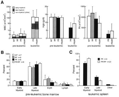
Figure 2.
Hematologic analysis of young transgenic animals. (A) Comparison of peripheral blood counts. Blood was obtained from six cohorts of healthy age-matched, genotyped littermates at 2–9 months of age. Values represent the mean ± SD of total WBC (expressed as cells/mm3), hemoglobin (Hgb, expressed as gm/dl), and platelets (Plt, expressed as Plt × 103/mm3). PR and PR/RP leukemic animals (n = 4 for each) were also analyzed. (B) Comparison of bone marrow differentials from healthy young animals. Differential counts of at least 150 cells were performed by two independent observers blinded to genotypes. Values reflect the mean ± SD, indicated by error bars. Early myeloid cells include blasts, promyelocytes, and myelocytes, and late myeloid cells include metamyelocytes, band, and neutrophils. “Eryth” includes all erythroid precursors. (C) Comparison of differential counts from the leukemic spleens of PR vs. PR/RP transgenic mice. More than 95% of the cells in all spleens were myeloid. Early myeloid cells and late myeloid cells were defined as described above. The percentage of early myeloid cells is significantly higher (P < 0.05) in the PR/RP-derived spleens.

