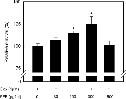
Fig. 1.
Cytoprotective activities of SFE on Dox-induced cytotoxicity in H9c2 cardiomyocytes. After 24 h exposure to 1 μM Dox, cells were incubated with SFE for a further 24 h and then assessed for cytotoxicity using the SRB assay. Cell survival is represented by a percentage of cell viability compared with the Dox-treated control. The concentration of SFE was expressed as a gallic acid equivalent (GAE). *p < 0.05 compared with the Dox-treated control

