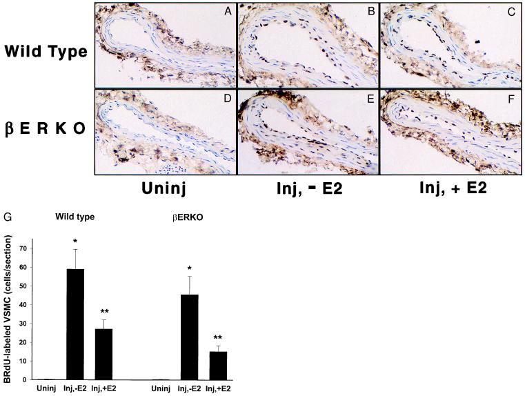
Figure 4.
Medial smooth muscle cell proliferation in uninjured and injured w.t. and βERKO mouse carotid arteries treated with vehicle or E2. (A–F) Representative segments of BrdU-stained carotid artery sections are shown (×400). The number of BrdU-positive smooth muscle cells for each section is listed. All BrdU counts were made for complete sections. (A) Wild-type, uninjured (Uninj; 0 cells). (B) Wild-type injured, vehicle-treated (Inj, −E2; 58 cells). (C) Wild-type, injured, E2-treated (Inj, +E2; 29 cells). (D) βERKO (Uninj; 0 cells). (E) βERKO (Inj, −E2; 45 cells). (F) βERKO (Inj, + E2; 17 cells). (G) Summary medial VSMC BrdU labeling data for all w.t. and βERKO mice. The number of BrdU-labeled cells did not differ between the w.t. and βERKO mice in the injured, vehicle-treated groups (59 ± 10 cells per section vs. 46 ± 10 cells per section, respectively) or in the injured E2-treated groups (27 ± 5 cells per section vs. 15 ± 3 cells per section, respectively). *, P < 0.05, compared with both uninjured, and injured, estrogen-treated groups within the same genotype; **, P < 0.05, compared with uninjured within the same genotype.

