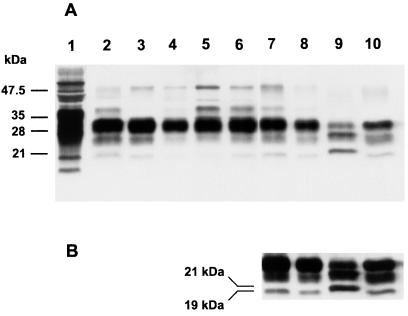
Figure 4.
Characterization of PrPSc fragments by immunoblot. Western blots of PrP in brains from Tg mice inoculated with BSE, nvCJD, and sporadic CJD (sCJD) prions. Lane 1, undigested PG31/90 BSE brain. Lane 2, PG31/90 BSE brain control. Lanes 3 and 4, Tg(BoPrP)4125/Prnp0/0 mice clinically ill after inoculation with BSE inocula PG31/90 and GJ248/85, respectively. Lanes 5 and 6, Tg(BoPrP)4125/Prnp0/0 mice clinically ill after inoculation with PG31/90 and GJ248/85, respectively, after a single serial passage in Tg(BoPrP)4125/Prnp0/0 mice. Lanes 7 and 8, Tg(BoPrP)4125/Prnp0/0 mice clinically ill after inoculation with prions from nvCJD patient RU96/02. Lane 9, sCJD, patient RG. Lane 10, nvCJD patient RU96/02. A is an exposure selected to clearly show differences in the PrP glycoform ratios. B is a longer exposure of lanes 7–10, selected to make the lower molecular weight unglycosylated fragment more evident.

