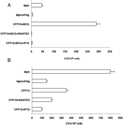
Figure 6. T cells co-cultured with or recruited by RANTES or IP-10 conditioned CFP10-DCs mediate clearance of BCG from macrophages.
For A, BCG infected CFP10-DCs (CFP10), conditioned or not with either 25 ng/ml RANTES or IP-10 were co-cultured for 48h with BCG primed T cells. From this, T cells were enriched and cultured with BCG infected macrophages (Mph) for 48h. A separate group wherein macrophages conditioned with 2 ng/ml IFN-γ (IFN-g) for 4h prior to infection with BCG was also included as a control. For B, T cells migrated into the lower chamber of a transwell apparatus in response to supernatants of BCG infected CFP-DCs, conditioned or not with 25 ng/ml RANTES or IP-10, were cultured with BCG infected macrophages (Mph) for 48h. A separate group wherein macrophages conditioned with 2 ng/ml IFN-γ (IFN-g) for 4h prior to infection with BCG was also included as a control. Cells from both Panels were lysed and plated in serial dilutions on 7H11 agar plates. CFU were counted 2–3 week later. Data are the mean±s.d. of three experiments. For A, P<0.002 (Mph vs CFP10+BCG+RANTES), P<0.004 (Mph vs CFP10+BCG+IP10). For B, P<0.02 (Mph vs CFP10+BCG+RANTES), P<0.01 (Mph vs CFP10+BCG+IP10).

