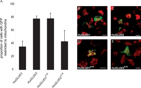
Figure 5. Subcellular Localization of GLUD1 and GLUD2 Proteins With Wild Type and Mutant MTSs, respectively.
(A) Proportion of transfected HeLa cells with GLUD-GFP signals detected only in mitochondria. HuGLUD2K7E shows significantly lower mitochondrial localization specificity than HuGLUD2 and HuGLUD1E7K (P<0.01, Tukey's Post Hoc test). (B)–(E) HeLa cells transfected with wild type and mutant GLUD proteins (mitochondria labeled with MitoTracker, red). Scale bars = 10 µm. Unmerged images are shown in Figure S5.

