
Figure 3.
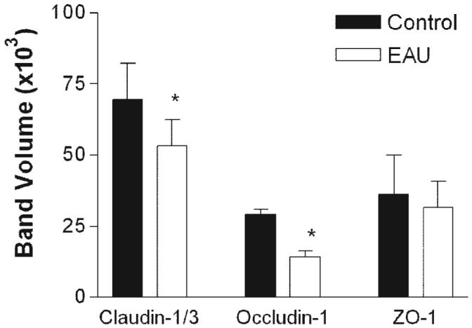
Western blot analysis for TJ proteins in retinas from normal and EAU mice. Retinas were dissected from the choroids-RPE tissue in normal or EAU mice (C57BL/6 milder disease) and proteins extracted for Western blot analysis with claudin-1/3, occludin-1, and ZO-1. Data shown are expressed as the mean ± SEM. n = 3 mice, *P < 0.05 Student’s t-test.