
Figure 5.
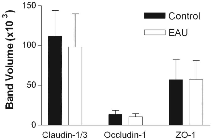
Quantification of results in a Western blot analysis of TJ proteins in RPE cells from normal and EAU mice. RPE-choroid tissues were dissected from normal or EAU mice (C57BL/6 milder disease) and proteins extracted for Western blot analysis with claudin-1/3, occludin-1, and ZO-1. Data are expressed as the mean ± SEM; n = 3 mice.