
Figure 6.
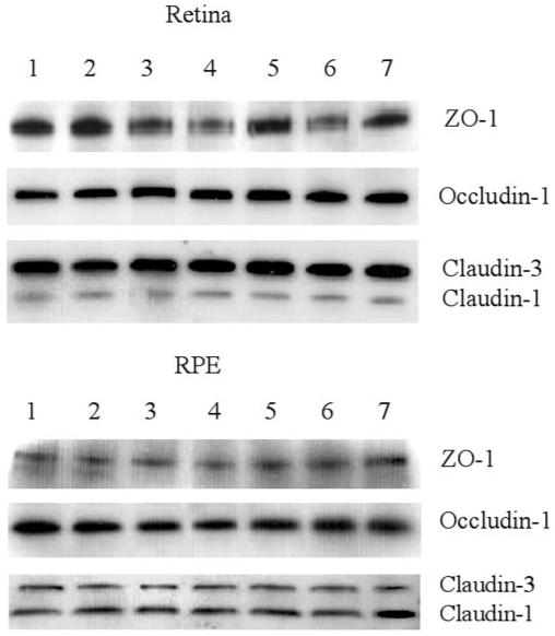
Western blot analysis of ex vivo retinas and RPE cells for TJ proteins ZO-1, occludin-1, and claudin-1/3 after cytokine or chemokine treatment. Retinal and choroid-RPE tissues were dissected from normal mouse eyes and incubated for 24 hours in the presence or absence of different cytokines or chemokines. Proteins were extracted for Western blot analysis. Row 1, control; row 2, IFN-γ; row 3, TNF-α; row 4, IL-1β; row 5, MCP-1; row 6, MIP-1α; and row 7, RANTES. Data are representative of three experiments. Bands representing either claudin-1 (20 kDa) or claudin-3 (22 kDa) were differentiated in blots, by using specific claudin-1 and claudin-3 antibodies that became available during the period of the study (data not shown).