
Figure 7.
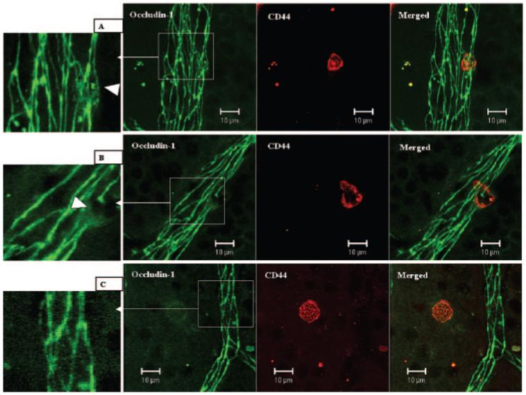
Changes of occludin-1 in retinal venules during leukocyte adhesion and transendothelial cell migration. Day 9 pi EAU mice were gently perfused with PBS. Retinal wholemounts were double stained with anti-occludin-1 (FITC) and anti-CD44 (R-PE). (A) One CD44+ leukocyte adhering to a retinal venule. TJ protein occludin-1 is diffuse in the adhering area (inset, arrowhead). (B) One CD44+ leukocyte was in the process of transendothelial cell migration. TJ protein occludin-1 was absent in the transmigration area and diffuse in the adjacent area (inset, arrowhead). (C) One CD44+ cells had migrated into the tissue, and there was no occludin-1 disruption in surrounding vessels. Images are reconstructions from a series of Z-stacks (15-40-μm thickness). Bars, 10 μm.