
Figure 8.
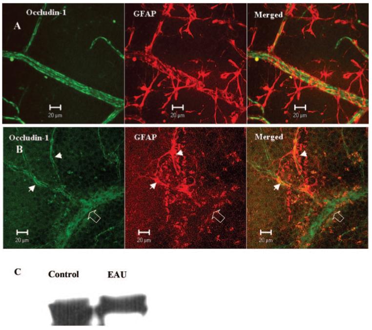
Occludin-1 and GFAP immunoreactivity in normal and EAU mouse retina. Retinal wholemounts from a normal B10R.III mouse (A) and a day 12 pi EAU mouse (B) were stained with anti-occludin-1 and anti-GFAP. Samples were observed by confocal microscopy. Note the total loss of GFAP staining in the vessel segment with occludin-1 disruption (diffuse occludin-1 staining, open arrow) and the preservation of occludin-1 in capillaries where GFAP astrocytes are maintained intact (arrowheads). Images shown are reconstructions from a series of Z-stacks (40-μm thickness). (C) Western blot of retinal tissue from control and EAU mice showing reduced GFAP protein expression in EAU retina.