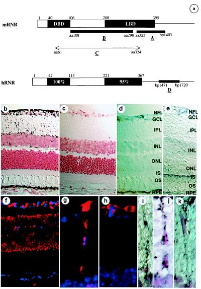
Figure 2.
In situ hybridization on albino mouse and rhesus monkey retina by using RNR-specific probes. (a) Portions of mRNR and hRNR used for generating the in situ probes. (b–e and i–k) In situ hybridizations on albino mouse retina with chromogenic detection and counterstaining with neutral red; hybridization signal is seen as black and retinal cell nuclei are seen as red. (b) Antisense probe A. (c) Sense probe A. (d) Antisense probe B. (e) Antisense ABCR probe. (i–k) Higher-power views of cells having morphology of Müller glia in the inner nuclear layer/inner plexiform layer (IPL). (f) In situ hybridization of antisense probe D on rhesus monkey retina; in situ signal is detected by using coumarin fluorescence and is seen as blue; cell nuclei are counterstained by using propidium iodide and are seen as red. (g–h) Glial fibrillary acidic protein immunohistochemistry on mouse eye sections adjacent to those used in b–e, seen as red fluorescence; cell nuclei are seen as blue. NFL, nerve fiber layer; GCL, ganglion cell layer; INL, inner nuclear layer; ONL, outer nuclear layer; IS, inner segments of photoreceptors; OS, outer segments of photoreceptors; RPE, retinal pigment epithelium.

