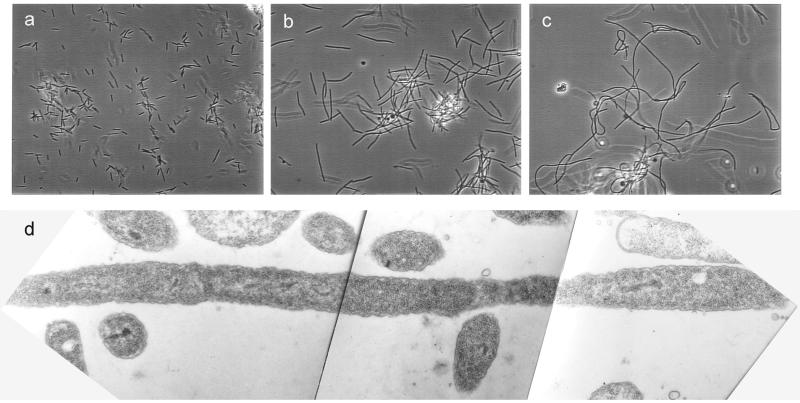
Figure 1.
Effect of cephalexin on M. xanthus. Phase-contrast images of wt M. xanthus cells (DK1622) grown in CYE without cephalexin (a), with 100 μM cephalexin after 6 hr (b), and with 100 μM cephalexin after 12 hr (c). Pictures were taken through a ×32 objective lens. (d) Electron microscopy analysis of DK1622 myxo-filaments grown in CYE with 100 μM cephalexin for 12 hr (×14,000).

