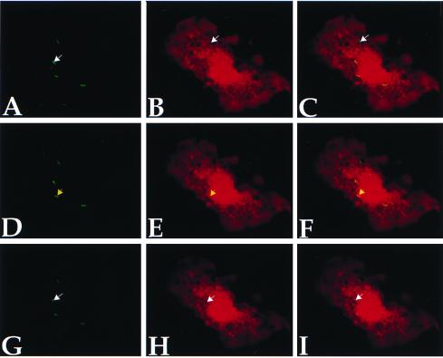
Figure 2.
Signal colocalization is not a plane of section artifact. BCG-GFP-infected macrophages were microinjected with 40-kDa-sized Texas Red-tagged dextrans and immediately were visualized by confocal microscopy. Successive optical sections were collected along the z axis, with a step of 0.9 μm. Three optical planes taken from the substratum up—A–C, D–F, and G–I—are shown. A, D, and G show fluorescence in the green channel only, B, E, and H show fluorescence in the red channel only, and C, F, and I are merged for RGB color. Numerous bacilli (A, white arrow) remained within clearly discernable vesicles impenetrable to the dextrans (B, white arrow), thereby precluding signal colocalization. Occasionally, yellow bacilli seemingly within compartments accessible to the dye appeared within an impermeable vesicle only after evaluation of several successive optical sections (compare bacteria in I, white arrow, with C). A significant number of BCG-GFP, however, facilitated dextran signal colocalization at every plane of section (D–F, yellow arrows).

