
Figure 3. CaM immunoreactivities at the calyces of Held terminal at P8 and P14.
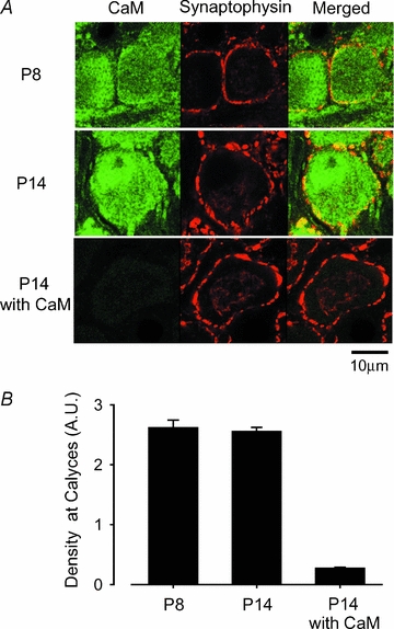
A, CaM immunoreactivity (left column, labelled green with Alexa fluor 488), synaptophysin immunoreactivity (middle column, labelled red with Alexa fluor 568), and their overlap (right column, yellow). Bottom panel in the left column (P14 with CaM) shows the background after absorbing the primary antibodies with CaM protein. B, densitometric measurements of CaM immunofluorescence intensity in the regions overlapped with synaptophysin immunofluorescence signals at P8 and P14, and the background intensity after antibody absorption at P14.