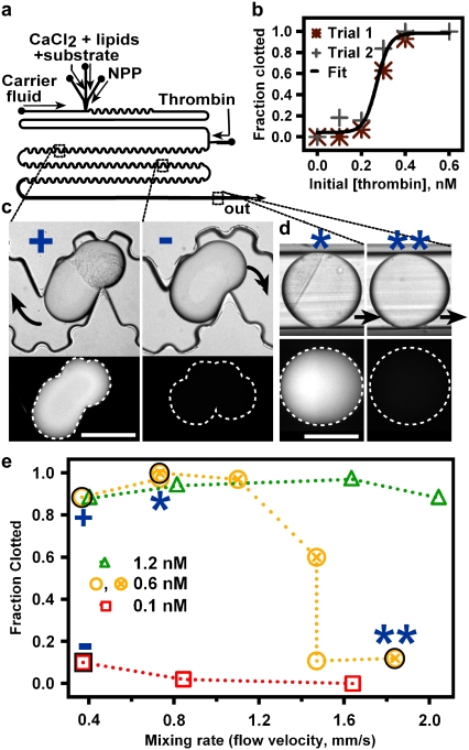
FIGURE 2.
Rapid mixing prevents clotting in human NPP near the reaction threshold. (a) Schematic of the device used to merge droplets of an activator, C (thrombin), with plugs of recalcified, relipidated NPP and a fluorogenic substrate for thrombin. (b) In a straight, smooth channel, the fraction of plugs (n ≥ 30) that clot increased from 0 to 1 as the concentration of thrombin, [C]i, injected into the NPP plug increased from 0.0 to 0.6 nM (2 parts 0.0–0.6 nM thrombin/6 parts recalcified, relipidated NPP with substrate, by volume). Black line shows a sigmoidal curve drawn to guide the eye. (c and d) Wide-field fluorescence and brightfield microphotographs of plugs with clotted (clumps of particles, bright fluorescence) and unclotted (no clumps, no fluorescence) NPP flowing through a microfluidic device after merging with 0.6 nM (+, *, **) or 0.1 nM (−) [C]i. Blue symbols correspond to those in e. Black arrows indicate direction of fluid flow. Scale bars, 300 μm. (c) Slowly flowing (0.37 mm/s) plugs are visible in the channel 20 min after merging. Those receiving 0.6 nM (+) and 0.1 nM (−) [C]i all clotted or remained unclotted, respectively. (d) Faster flowing plugs were monitored in Teflon tubing 20 min after merging. Those receiving [C]i ≈ [C]i,crit either clotted (*) or did not (**), depending on the mixing rate. (e) In a winding, bumpy channel, the fraction of plugs that clotted within 20 min decreased with increasing mixing rate when [C]i ≈ [C]i,crit (0.6 nM thrombin).

