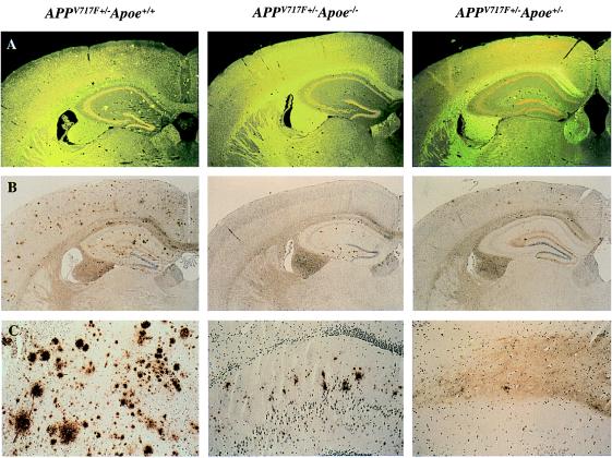
Figure 1.
Lack of apoE reduces both Aβ immunoreactive as well as amyloid deposits in a transgenic mouse model of AD. In 21/22-mo-old APPV717+/− TG mice wild-type for Apoe (APPV717F+/− Apoe+/+), numerous thioflavine-S-fluorescent (A) and Aβ immunoreactive deposits (B and C) are evident in both the hippocampus and the cerebral cortex. By contrast, in 21/22-mo-old APPV717F+/− Apoe−/− mice, no thioflavine-S-fluorescent Aβ deposits are observed in any brain region. Furthermore, Aβ-immunoreactive deposits are confined to the hippocampus. APPV717F+/− Apoe+/− mice had an essentially intermediate level of both thioflavine-S-fluorescent and Aβ immunoreactive deposits. Neither thioflavine-S-fluorescent nor Aβ-immunoreactive deposits were found in APPV717F−/− Apoe+/+ mice. The photomicrographs are from representative sections of each of the genotypes indicated. (Original magnification, A and B ×9, C ×35.)

