
Figure 5.
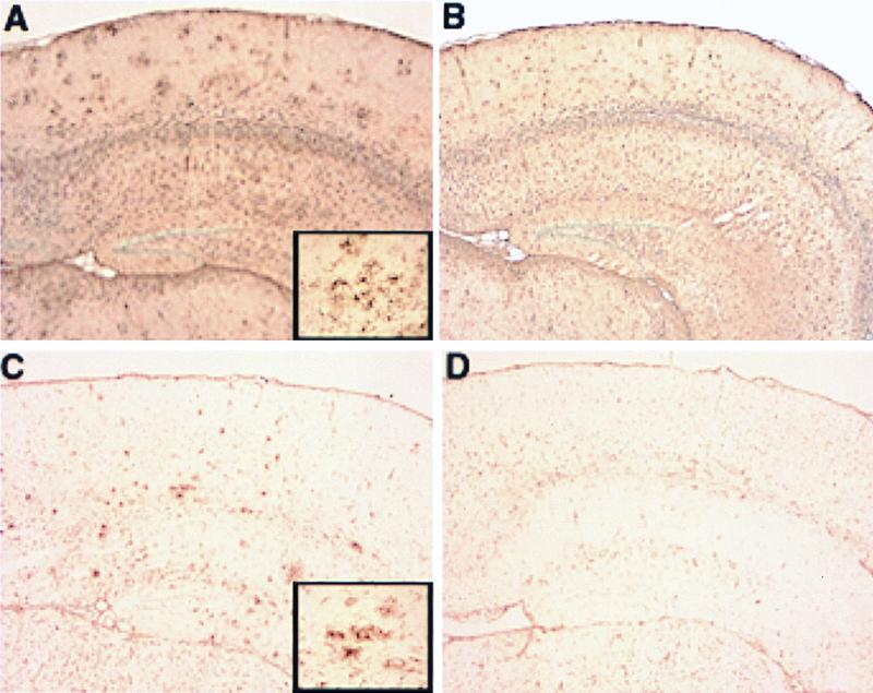
Reduced glial activation in APPV717F+/− Apoe−/− TG mice. Glial activation was evaluated by GFAP immunohistochemistry (A and B) and tomato lectin histochemistry (C and D) on paraffin-embedded brain sections from 21/22-mo-old APPV717F+/− Apoe+/+ and APPV717F+/− Apoe−/− mice (see Materials and Methods for details). A range of glial staining for each genotype analyzed was apparent with an increase in GFAP immunoreactivity and lectin staining in the superficial layers of the cerebral cortex and hippocampus in APPV717F+/− Apoe+/+ mice (A and C, respectively) compared with APPV717F+/− Apoe−/− mice (B and D, respectively). Note clusters of intensely stained astrocytes (A, Inset) and microglia (C, Inset), which are more prominent in APPV717F+/− Apoe+/+ than in APPV717F+/− Apoe−/− mice. (×5; Inset, ×40.)