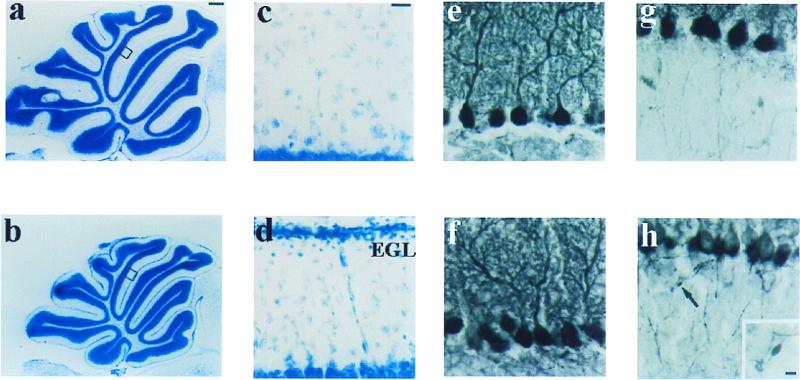
Figure 2.
Histological and immunohistochemical analyses of brain in wt and α1A−/− mouse at P21. (a and b) Toluidine blue-stained sagittal sections of cerebellar vermis. (c and d) Higher magnification of areas in a and b. The mutant showed the persistence of an external granule cell layer, indicating a delay or deficit in granule cell migration (d). (e– h) Cerebellar cortex immunostained with anticalbindin antibody. (e and f) Dendritic trees of Purkinje cells. (g and h) Axons of Purkinje cells in granule cell layer. Arrow indicates focal axonal swellings that frequently are found in α1A−/− (h Inset). EGL, external granule cell layer. [Bars = 400 μm (a and b), 25 μm (c– h), and 5 μm (h Inset).]

