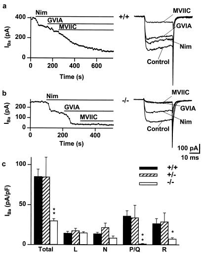
Figure 3.
P/Q-type calcium channels are absent and R-type calcium channels are diminished in α1A−/− cerebellar granule cells. (a) Peak IBa, activated by 30-ms depolarizations from –90 mV to –10 mV every 10 s, is plotted against time for an α1A+/+ neuron. The total current was reduced by the application of nimodipine (10 μM), ω-CTx-GVIA (1 μM), and ω-CTx-MVIIC (5 μM), which blocked the L-, N-, and P/Q-type components of the current, respectively. Representative traces are shown to the right of the graph. Ba2+ (10 mM) as charge carrier. (b) Peak IBa, activated as in a, is plotted against time for an α1A−/− neuron. (c) Histogram of total and individual channel type current density in α1A+/+ (solid), α1A+/− (hatched), and α1A−/− (open) granule cells. The total current density is reduced in α1A−/− neurons; this is accounted for by the absence of the P/Q-type channels and the reduction of R-type current. **, P < 0.002, *, P < 0.05.

