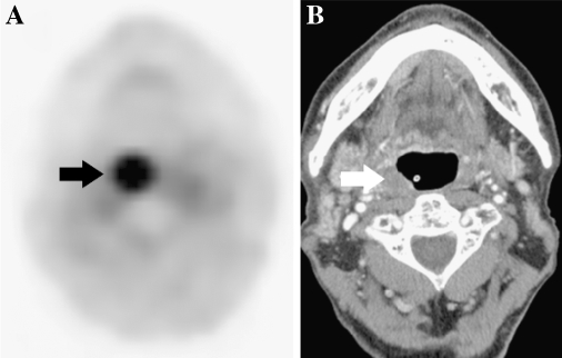
Fig. 3.
Discrepancy between post-CRT PET and CT findings in a 78-year-old male with OPC of the right lateral wall, tonsil, treated with concurrent CRT. Local recurrence was confirmed histologically about 6 months later. A contrast-enhanced CT scan resulted in false-negative findings. a In post-CRT PET, strong uptake tracer was seen the right lateral wall (arrow). b Post-CRT axial CT scan showed normal to slight hypertrophy of the tonsil without contrast enhancement (arrow), and histological specimen immediately after the end of CRT indicated no evidence of disease.

