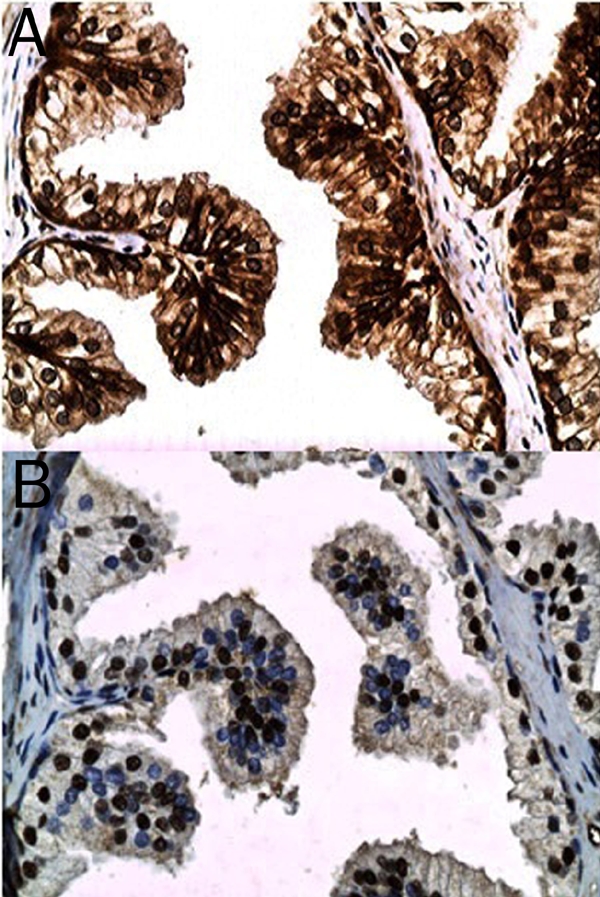
Figure 1.

Non-neoplastic glandular epithelium from a case of benign prostatic hypertrophy illustrating the relatively moderate intensity DAB (brown) chromogenic signal for mTOR (phosphorylated at serine 2448) in the plasmalemmal and cytoplasmic compartments of the luminal epithelium and the relatively strong signal in the basal cell layer (A). Variable chromogenic signal for p70S6K (phosphorylated on threonine 389) is noted in the nuclei of the glandular epithelium (B) (original magnification, ×400).
