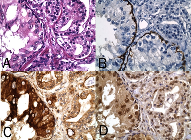
Figure 3.

HGPIN and companionate prostate cancer on H&E (A) and with the HGPIN confirmed by IHC staining for high molecular weight cytokeratin of a basal cell layer (B). Note relatively stronger DAB (brown) chromogenic signal for cytoplasmic/ plasmalemmal expression of p-mTOR (Ser 2448) (C) and for nuclear expression of p-p70S6K (Thr 389) (D) in HGPIN (original magnification, ×400).
