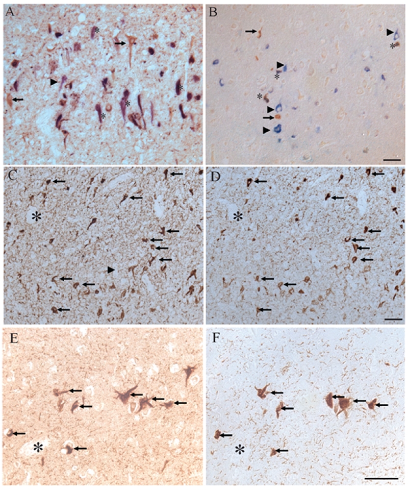
Figure 4.

Using double-label immunocytochemistry, localization of ppRb(S807), stained brown, was compared with PHF-1 (blue, A) and 12E8 (B). Neurons containing both phosphorylated tau and ppRb(S807) were marked by *. Neurons containing only ppRb(S807) (arrows), or only phosphorylated tau (arrowheads) were also marked. In adjacent sections of other hippocampal regions in AD, there is striking overlap of ppRb(S807) with PHF-1 (C, D respectively) as well as with the small number of NFT in an aged control case (E, F). Scale bar = 50 μm.
