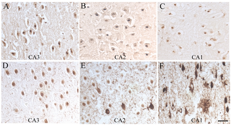
Figure 6.

Nuclear localization of ppRb is readily detected in sections of AD and control following pretreatment of the tissue sections with trypsin. In this figure, antisera to ppRb(S807) recognize nuclei in pyramidal neurons in a section from a control case, throughout the CA3, CA2 and CA1 regions (A, B, C). Conversely, in a single section from a representative AD case, pyramidal neuronal nuclei are positive in the CA3 area (D), while nuclei and a few NFT stain in the CA2 (E), and pathological structures are positive in the CA1, while the nuclei remain unstained (F). Scale bar = 50 μm.
