
Figure 2.
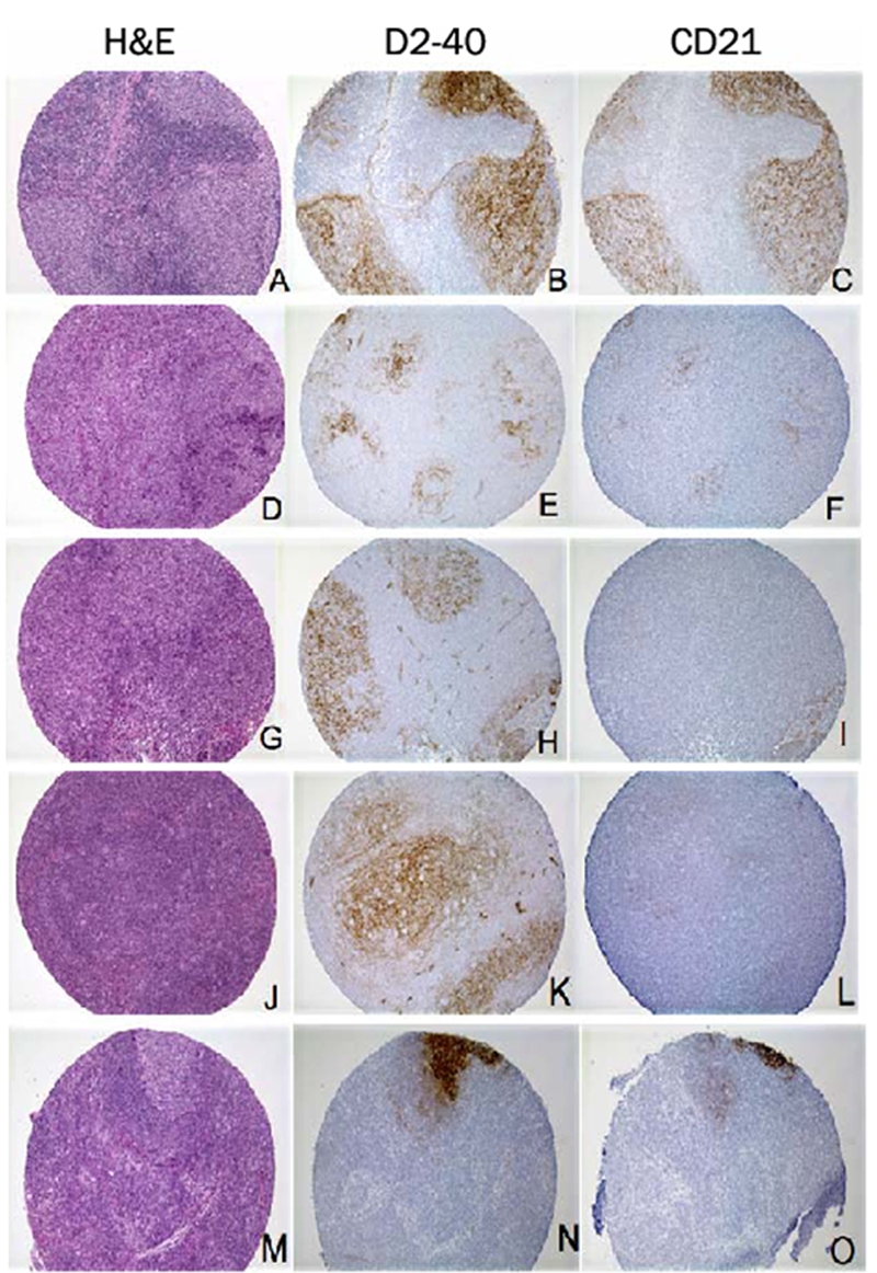
D2-40 immunohistochemical staining pattern in lymphomas. A-I. Follicular lymphoma: H&E staining reveals tightly apposed follicles (A, D and G). D2-40 (B) and CD21 (C) show identical staining patterns and intensity of the FDC meshworks. D2-40 (E) immunostaining is much stronger in FDC meshworks than CD21 (F). D2-40 (H) is positive in this disrupted FDC meshwork, while CD21 (I) fails to highlight the same FDC meshwork. J-L. Nodular lymphocyte predominant Hodgkin lymphoma: H&E staining reveals markedly expanded follicle (J). D2-40 (K) immunostaining is much stronger in the markedly expanded FDC meshworks than CD21 (L). M-O. Diffuse large B-cell lymphoma (DLBCL): H&E staining reveals diffuse area with focal residual germinal center (M). D2-40 (N) immunostaining is much stronger in residual FDC meshwork than CD21 (O). (Original magnification: A-O, ×100)