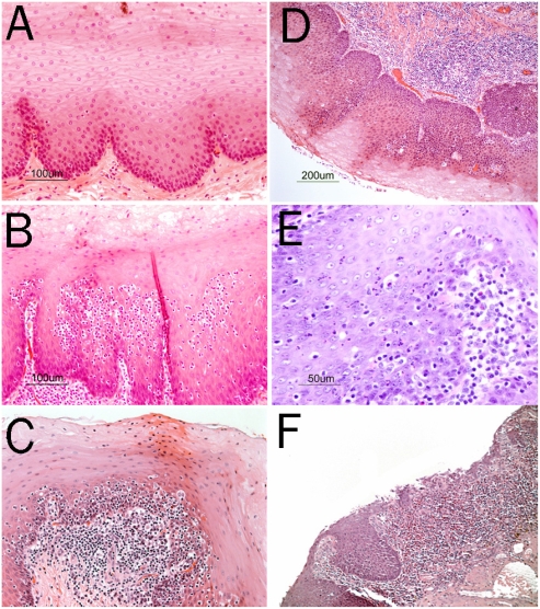
Figure 1.
A. Normal squamous epithelium of the esophagus in a baboon (H&E, original magnification 20×). B and C. Lymphocytic esophagitis in baboons showing infiltration of the squamous epithelium of the esophagus around the papillae by a high numbers of lymphocytes as well as chronic inflammation in the lamina propria (H&E, original magnification 20×). D. Grade 1 reflux esophagitis in a baboon (D) with tall papillae and basal-cell proliferation and few intraepithelial lymphocytes. Chronic inflammation in the lamina propria is also seen (H&E, original magnification 10×). Grade 2 reflux esophagitis in a baboon (E) with intraepithelial granulocytes and chronic inflammation in the lamina propria (H&E, original magnification 20×). Grade 3 reflux esophagitis in a baboon (F) with mucosal ulceration and severe chronic inflammation in the lamina propria (H&E, original magnification 20×).

