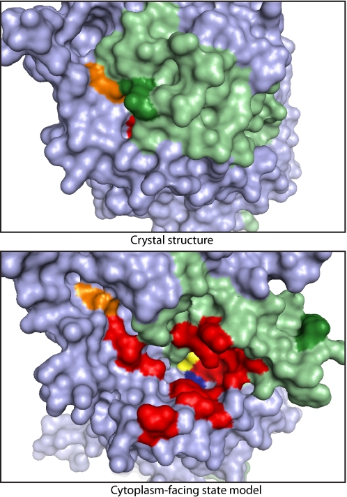
Fig. 6.
Molecular surface of LeuT, viewed from the cytoplasmic side of the membrane. (Upper) X-ray crystal structure. (Lower) Model of the cytoplasm-facing state. The bundle is shaded light green and the remainder of the protein light blue. The surface of leucine (yellow), Na2 (blue), the Arg-5 (green), and Asp-369 (orange) ion pair and residues corresponding to those found to be accessible in SERT (red and orange) are highlighted.

